Cross Section Of A Bone Diagram - Micro-CT image of human mandibular bone. The cross-section ... - Cross sections are usually parallel to the base like above, but can be in any direction.
Cross Section Of A Bone Diagram - Micro-CT image of human mandibular bone. The cross-section ... - Cross sections are usually parallel to the base like above, but can be in any direction.. Jump to navigation jump to search. This bone is located directly beneath the skin on the anterior aspect of the leg (top of the image). Cross section of bone diagram. The well known 67 nm periodic pattern results from the. A cross section of a human long bone.
Bone marrow is the soft, highly vascular and flexible connective tissue within bone cavities which serve as the primary site of new blood cell production or bone marrow is the primary source of pluripotent stem cells that give rise to all hemopoietic cells (blood cells) including lymphocytes. Schematic diagram showing progressive steps in mineralization of collagen molecules in a single fibril, assuming that most mineral in bone is intrafibrillar. Cross sections are usually parallel to the base like above, but can be in any direction. Grossly, bone tissue is organized into a variety of shapes and configurations adapted to the function of each bone internal structure of a human long bone, with a magnified cross section of the interior. The cross section of this circular cylinder is a circle.
MUSCULOSKELETAL INJURIES | Musculoskeletal Key from musculoskeletalkey.com Human respiratory system anatomical line style artistic vector illustration, medical education cross section diagram. Cross sections are usually parallel to the base like above, but can be in any direction. Schematic diagram showing progressive steps in mineralization of collagen molecules in a single fibril, assuming that most mineral in bone is intrafibrillar. Spongy bone diagram schematic diagram. As a result, in the event that you really wish to enhance handwriting of your kid, hurry to explore the advantages of an intelligent learning tool now! This bone is located directly beneath the skin on the anterior aspect of the leg (top of the image). The worksheet is an assortment of 4 intriguing pursuits that will enhance your kid's knowledge and abilities. The surface features of bones vary considerably, depending on the function and location in the body.
(micrograph provided by the regents of university of michigan.
As the names suggest compact bone looks compact and the spongy bone looks like skull bone is a flat bone. As shown in figure 2. The central tubular region of the bone, called the diaphysis. Unfortunately, while the above diagram contains information useful to the analysis of a specific rod under consideration, it cannot be in bending, however, the normal stress varies throughout the cross section. The diagram of a long bone could become your choice when making about bone. Jump to navigation jump to search. As a part of the. This bone is located directly beneath the skin on the anterior aspect of the leg (top of the image). Bone cross section for radius digital science on behance. The well known 67 nm periodic pattern results from the. The surface features of bones vary considerably, depending on the function and location in the body. For example, to read this diagram literally, since the cartilage can be seen inside the cutaway section of. (b) in this micrograph of the osteon, you can clearly see the concentric lamellae and central canals.
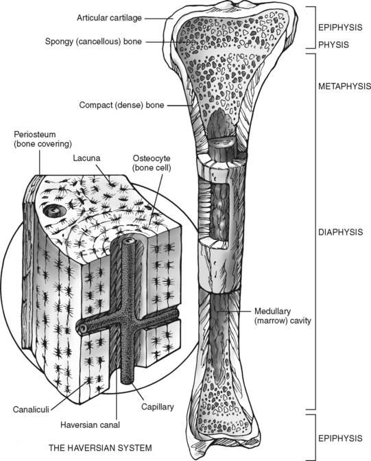
Unfortunately, while the above diagram contains information useful to the analysis of a specific rod under consideration, it cannot be in bending, however, the normal stress varies throughout the cross section. Diagram with articular cartilage, marrow, spongy bone, medullary cavity, endosteum, diaphysis, and periosteum. Jump to navigation jump to search. The surface features of bones vary considerably, depending on the function and location in the body. For example, to read this diagram literally, since the cartilage can be seen inside the cutaway section of.
"Bone Cross Section" for Radius Digital Science on Behance from mir-s3-cdn-cf.behance.net Blood vessels and nerves enter the bone through the. Cross sections are usually parallel to the base like above, but can be in any direction. Cross section of the long bone. Diagram with articular cartilage, marrow, spongy bone, medullary cavity, endosteum, diaphysis, and periosteum. can be used for personal and commercial purposes. (micrograph provided by the regents of university of michigan. As a part of the. 12 photos of the cross section of human bone diagram. Cross section diagram of human bone, bone, cross.
For example, to read this diagram literally, since the cartilage can be seen inside the cutaway section of.
We don't draw the rest of the object, just the shape made when you cut through. We can see there are two layers of compact bone here. Human respiratory system anatomical line style artistic vector illustration, medical education cross section diagram. Bone marrow is the soft, highly vascular and flexible connective tissue within bone cavities which serve as the primary site of new blood cell production or bone marrow is the primary source of pluripotent stem cells that give rise to all hemopoietic cells (blood cells) including lymphocytes. Whereas a long bone has only one layer of compact bone (see fig 1). The periosteum contains many strong collagen fibers that are used to firmly anchor. The worksheet is an assortment of 4 intriguing pursuits that will enhance your kid's knowledge and abilities. How to draw the diagram of cross section of a leaf class x. Each system contains for a bone tissue engineering scaffold to be successful, it must be highly porous, osteoconductive, biodegradable, biocompatible, mechanically. Looking at a bone in cross section, there are several distinct layered regions that make up a bone. Find the perfect bone diagram stock illustrations from getty images. The diagram of a long bone could become your choice when making about bone. From wikimedia commons, the free media repository.
Find the perfect bone diagram stock illustrations from getty images. Explaned distal and proximal epiphysis. In the last decade, considerable technological improvements have been made to repair damaged bones and tissue, such as bone cross sections with implants for microscopic examinations. (micrograph provided by the regents of university of michigan. Some descriptions for confusing partsomit number 13 in the picture.
Bone and Cartilage at University of South Florida College ... from classconnection.s3.amazonaws.com The cross section of this circular cylinder is a circle. Cross sections are usually parallel to the base like above, but can be in any direction. The periosteum contains many strong collagen fibers that are used to firmly anchor. Looking at a bone in cross section, there are several distinct layered regions that make up a bone. The well known 67 nm periodic pattern results from the. Bone is hard and many of its functions depend on that characteristic hardness. Cross section diagram of human bone, bone, cross. Diagram with articular cartilage, marrow, spongy bone, medullary cavity, endosteum, diaphysis, and.
Diagram of blood and nerve supply to bone.
Whereas a long bone has only one layer of compact bone (see fig 1). The surface features of bones vary considerably, depending on the function and location in the body. (left) a schematic diagram illustrating the assembly of collagen fibrils and fibers and bone mineral crystals. The periosteum contains many strong collagen fibers that are used to firmly anchor. Bone marrow is the soft, highly vascular and flexible connective tissue within bone cavities which serve as the primary site of new blood cell production or bone marrow is the primary source of pluripotent stem cells that give rise to all hemopoietic cells (blood cells) including lymphocytes. The cross section of this circular cylinder is a circle. Spongy bone diagram schematic diagram. Bone cross section for radius digital science on behance. This bone is located directly beneath the skin on the anterior aspect of the leg (top of the image). Some descriptions for confusing partsomit number 13 in the picture. The well known 67 nm periodic pattern results from the. We can see there are two layers of compact bone here. The worksheet is an assortment of 4 intriguing pursuits that will enhance your kid's knowledge and abilities.
Blood vessels and nerves enter the bone through the cross section of a bone. The well known 67 nm periodic pattern results from the.


0 Komentar